Pearson Edexcel International GCSE in Biology · 4BI1 · Free · no account needed
The Alimentary Canal
One long tube whose contents are still outside you, and the muscular squeeze that moves them along it.
Topic 2 · Structures and functions in living organisms — one of 34 lessons in this topic, and one of 68 in Biology.
This lesson ends with William Beaumont. Learn the Alimentary Canal, teach it, then defend it against William Beaumont.
The definition this lesson teaches, word for word — every Biology definition.
- Peristalsis
- Waves of muscular contraction that move food along the alimentary canal, produced by circular and longitudinal muscles in the gut wall contracting and relaxing alternately as an antagonistic pair.
What this lesson covers in the specification
Incandio is aligned to this specification. It is not published by, endorsed by or affiliated with Pearson, and it reproduces none of Pearson's wording — the statement numbers are given so you can check every lesson against your own copy.
- 2.27 — Structure and function of the human alimentary canal
- 2.28 — How peristalsis moves food through the gut
Specification section 2(e) Nutrition — also taught in Photosynthesis, What Limits Photosynthesis, The Leaf: Structure and Gas Exchange, A Balanced Diet.
1 · Understand it
No exam language yet. The only question this section answers is: do I actually understand what is happening?
Start with a fact that sounds like a trick and is not. The alimentary canal is a tube running from your mouth to your anus, and anything inside that tube is still outside your body. A swallowed coin is no more inside you than a coin held in your fist — it is in a channel that passes through you. Food only truly enters when it crosses the gut wall into the blood. Everything the gut does is aimed at that one moment, and holding this picture in your head makes the order of the organs obvious rather than something to memorise.
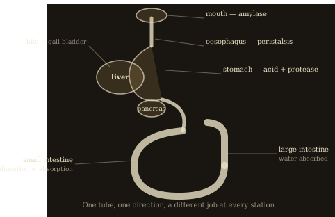
Why the organs come in the order they do
- MOUTH: teeth cut and grind the food into smaller pieces. This is physical, not chemical — it does not change the molecules, it makes many small pieces where there was one large one, so enzymes have far more surface to work on. Saliva adds amylase and lubricates the lump for swallowing.
- OESOPHAGUS: no digestion here at all. It is a delivery pipe from the throat to the stomach.
- STOMACH: a muscular bag that churns the food and adds hydrochloric acid and protease. It holds the meal for hours, which is why one meal can last you a morning.
- SMALL INTESTINE: the longest section, and where the real work happens. The first part receives bile and pancreatic juice; digestion is completed here and the soluble products are absorbed into the blood.
- LARGE INTESTINE: what is left is mostly water and indigestible fibre. Water is absorbed back into the blood here, so what remains becomes progressively more solid.
- RECTUM and ANUS: the solid waste, faeces, is stored and then egested. Notice that this material was never absorbed, so it was never part of you — egestion is not excretion.
Now the movement. Food does not fall through you, and it is worth being sure of that: an astronaut in orbit can eat perfectly well, and you can swallow while standing on your head. Gravity has almost nothing to do with it. The gut wall carries two layers of muscle — one set running around the tube in rings, the other running along its length — and they work as an antagonistic pair, meaning that when one contracts the other relaxes.
One wave of peristalsis
- A ball of food, the bolus, is sitting in the tube.
- The circular muscles BEHIND the bolus contract, and the longitudinal muscles there relax. That section becomes narrow and long, squeezing the bolus forwards.
- At the same moment the circular muscles IN FRONT relax and the longitudinal muscles contract, so the tube ahead becomes wider and shorter — an open space for the bolus to be pushed into.
- The point of contraction then travels along the tube, so the squeeze follows the bolus rather than staying still.
- The result is a wave of narrowing that moves the contents steadily in one direction, whatever position the body is in.
This is also where fibre earns its place in the last lesson. The muscles can only push what they can grip. Contents with plenty of bulk are squeezed along efficiently; contents with very little are harder to move, the waves become less effective, and material stays in the large intestine longer — where more and more water is absorbed from it, making it harder still.
2 · Grade 9 Notes
A different job from the section above. You have already understood it; this is the precise set of things to LEARN — definitions to reproduce word for word, processes in order, equations with units, and the answers that score full marks.
Learn this definition · Peristalsis
Waves of muscular contraction that move food along the alimentary canal, produced by circular and longitudinal muscles in the gut wall contracting and relaxing alternately as an antagonistic pair.
The five words for what happens to food — examiners test these against each other
- Ingestion
- taking food into the body through the mouth
- Digestion
- breaking large insoluble molecules into small soluble ones, physically and then chemically by enzymes
- Absorption
- the movement of small soluble molecules through the gut wall into the blood
- Assimilation
- the use of absorbed molecules by the body's cells to build tissue or release energy
- Egestion
- removal of undigested food as faeces; it was never absorbed, so it is NOT excretion
Each part of the tube and the one thing it does
- Mouth
- teeth break food up physically, increasing surface area; saliva adds amylase and lubricates the bolus
- Oesophagus
- carries the bolus from mouth to stomach by peristalsis; no digestion occurs here
- Stomach
- churns food and secretes hydrochloric acid and protease; holds the meal for several hours
- Small intestine
- receives bile and pancreatic juice, completes digestion, and absorbs the soluble products into the blood
- Large intestine
- absorbs water from the remaining material back into the blood
- Rectum and anus
- store the faeces and egest them
One peristaltic wave — the sequence to write
- Circular muscles behind the bolus contract while longitudinal muscles there relax, narrowing the tube and squeezing the bolus forwards.
- Circular muscles in front of the bolus relax while longitudinal muscles there contract, widening the tube ahead of it.
- The point of contraction travels along the tube, so the squeeze follows the bolus.
- The contents are moved steadily in one direction, independently of gravity.
Model answer [4 marks]
Explain how peristalsis moves food along the oesophagus, and why a person can swallow while lying down. [4]
The circular muscles behind the bolus contract and the longitudinal muscles there relax, narrowing the tube and squeezing the bolus forwards. Ahead of it the circular muscles relax and the longitudinal muscles contract, widening the tube. This point of contraction travels along the oesophagus as a wave. Because the food is squeezed by muscle rather than falling, gravity is not needed and a person can swallow lying down.
Model answer [2 marks]
Explain the difference between egestion and excretion. [2]
Egestion is the removal of undigested food as faeces; this material passed through the gut without ever being absorbed, so it was never part of the body. Excretion is the removal of waste products made by the body's own metabolism, such as carbon dioxide and urea.
Mark-losing trap. The liver, gall bladder and pancreas are not part of the tube. They are organs beside it that pour secretions into it.
Mark-losing trap. Faeces are egested, not excreted. Anything never absorbed was never part of you, so it cannot be a metabolic waste product.
Mark-losing trap. Chewing is physical digestion. It increases surface area for enzymes; it does not break any chemical bonds.
Mark-losing trap. Peristalsis is not gravity and not a single muscle. Name BOTH muscle layers and say one contracts while the other relaxes.
3 · Prove it — the five questions
The five questions climb Grade 6 → Grade 7 → Grade 8 → Grade 9 → Grade 9 challenge, and are marked inside Incandio on your own device, by rule, with an authored diagnosis of the mistake you actually made. The mark schemes stay in the app so that the practice is worth doing; the questions themselves are here.
- Grade 6 · Identify [1 mark] — In which part of the alimentary canal is most water absorbed back into the blood?
- Grade 7 · State [1 mark] — Name the process by which undigested food is removed from the body as faeces.
- Grade 8 · Explain [4 marks] — Explain how a wave of peristalsis moves a bolus of food along the oesophagus. Select every statement that belongs in a full-mark explanation.
- Grade 9 · Calculate [3 marks] — An adult small intestine is about 6.0 m long with an internal diameter of about 3.0 cm. Treating it as a smooth cylinder, calculate the area of its internal surface in m². Give your answer to two significant figures.
- 9+ · Analyse [5 marks] — A patient's diet contains very little fibre. Over several months the material in their large intestine moves more and more slowly, and eventually becomes hard and difficult to egest. Select every statement that belongs in a full-mark explanation.
The people behind this science
Three ways into the same idea — the one who watched a living stomach for ten years, and the one who opened bodies and drew what was inside, and the one who compared the guts of hundreds of animals. Inside Incandio each of them answers knowing exactly which lesson you have just finished.
William Beaumont — the one who watched a living stomach for ten years
In 1822 a shotgun accident left the voyageur Alexis St. Martin with a permanent opening into his stomach, and Beaumont — the army surgeon who saved him — spent the next decade doing what no one before or since has done: watching a living human stomach digest. He lowered food in on silk strings, timed every change, and watched the churning movements this lesson describes. He is the closest anyone has come to seeing the inside of the tube you are labelling.
- “What did the inside of a working stomach actually look like?”
- “How did the stomach move the food that was inside it?”
- “What did the silk-string experiments tell you that no dissection could?”
- “How much of digestion did you think the stomach did — and what could your window not show you?”
- “Was it right to bind a poor man to your experiments by contract?”
Leonardo da Vinci — the one who opened bodies and drew what was inside
Leonardo dissected around thirty human bodies and drew the digestive organs more accurately than anyone had before, working out the arrangement by following the tube itself rather than by trusting the textbooks of his day. That is exactly the exercise this lesson asks of you — and he did it knowing that what the authorities said was inside a body often was not.
- “What surprised you most when you first traced the gut from one end to the other?”
- “How did you decide which organs were part of the tube and which merely sat beside it?”
- “Why did you trust your own dissections over the accepted texts?”
- “What can a drawing show about an organ that words cannot?”
Aristotle — the one who compared the guts of hundreds of animals
Aristotle dissected and described the digestive systems of a very large number of species and noticed that the shape of the gut tracks what the animal eats — long and complicated in animals that live on plants, short and simple in those that eat flesh. That is structure explained by function, and it is the reasoning this lesson asks you to apply to a single human tube.
- “Why is a plant-eater's gut so much longer than a meat-eater's?”
- “What does the shape of an animal's gut tell you about how it lives?”
- “How did you work out what an organ was for, without any way to test it?”
- “Which part of the digestive system puzzled you most, and why?”
Think you understand it?
Reading is the start. On Incandio a topic counts as mastered only once you have taught it and defended it — the two things a revision guide cannot ask of you.
Incandio this topic → Prove it. Teach it. Defend it. What does “Incandio it” mean?
- Prove it — five questions on the Alimentary Canal, marked on the spot, that tell you what was missing rather than just what was wrong.
- Teach it — Explain the Alimentary Canal in your own words to Ember, an AI pupil who does not have it yet and asks the awkward question. Understanding is credited; wording is not required.
- Defend it — Argue the Alimentary Canal against William Beaumont — three rounds, marked against descriptors you can read before you begin.
- Then the exam — read all 10 authored exam-style questions on this unit, each with its command word and its tariff, then have them marked on Incandio with the mistake you actually made named.
Completely free. No subscriptions, no paywalls, no account needed to start.
Carry on through the course
- Previous lesson: A Balanced Diet
- Next lesson: Digestive Enzymes and Bile
- Levels of Organisation — From organelle to organism — the five-step ladder that every later topic in biology is built on.
- Comparing Cells, and Stem Cells — What plant and animal cells share and where they differ — then why a cell specialising is one of the most powerful ideas in biology.
- Carbohydrates, Proteins and Lipids — The three big food molecules — what they are made of, what they are built from, and how to test for them.
- Enzymes: Catalysts With a Shape — Why a protein with a hole in it speeds up a reaction, and why heat destroys that ability permanently.
- Enzymes and pH — Why acid ruins one enzyme and is the only condition another will work in — and how to find an enzyme's best pH by experiment.
- All of Biology · Biology key terms · required practicals · Incandio Science · The 4BI1 specification